Multiple Choice
Identify the choice that best completes the
statement or answers the question.
|
|
|
The image below shows a lipid bilayer with a representation of a
particulat type of membrane transport. Labels 1 and 2 indicate the compartments and do not identify
any structures.
|
|
|
1.
|
Based on the above image which of the following is a incorrect statement?
a. | The concentration gradient of X would predict that X would flow from compartment 1 to
compartment 2. | b. | X is a polar molecule | c. | The membrane is selectively
permeable. | d. | X is being actively transported against it’s concentration
gradient. | e. | When only considering the concentration of X, the compartment labeled 2. is
hypertonic to compartment 1 |
|
|
|
The following image shows a concentration gradient with a selectively permeable
membrane. Assume the membrane is freely permeable by the solute shown in compartment 1.
|
|
|
2.
|
Assume the image above shows the system at time 0 minutes. If you return to the
system after 20 minutes, which best describes the results you would expect to find
a. | the system reached equilibrium | b. | some of the solute moved from compartment 1 to
compartment 2. | c. | equal amounts of solute is moving in either direction at any given
moment. | d. | relatively equal concentrations of solute on either side of the
membrane. | e. | all of the above are correct statements |
|
|
|
The following image is a U-tube with an unknown solute dissolved into water
divided by a selectively permeable membrane. Assume that the membrane impermeable to the solute but
freely permeable to water.
|
|
|
3.
|
Assume this sytem is shown at time 0 minutes. Whixh of the following statements
would best describe what happens to the system after 20 minutes?
a. | The solute would move until there is equal concentrations of solute on both side of
the membrane | b. | The water will move from side 2 to side 1. | c. | The water will move
from side 1 to side 2 | d. | The solute will diffuse from side 2 to side
1. | e. | Both the water and the solute will move from side 1 to side 2.
|
|
|
|
The following image shows a cell doing bulk transport.
|
|
|
4.
|
The above image is shows a type membrane transport. Which of the following
best describes this type of transport?
a. | Active transport | b. | Passive transport | c. | facilitated
diffusion | d. | receptor mediated endocytosis | e. | phagocytosis |
|
|
|
The diagram below shows a transmembrane protein (a protein embedded in the lipid
bilayer) that acts as a channel to transport molecules across the membrane. You should recognize the
parts of the lipid bilayer by comparing them to an earlier question which shows the membrane in the
same view. The boxed area highlights details of the protein chain that sits in the
membrane. Each “R” represents a separate sidechain and is labeled 1 through
5. Answer the following questions based on your understanding of the structure and
characteristics of amino acids, proteins, and the lipid bilayer.
|
|
|
5.
|
This channel protein, the potassium channel, facilitates the movement of
potassium ions across the cell membrane by what would be referred to as “facilitated
diffusion”. Which of the following would be true about the functions of this
protein?
a. | The protein is capable of moving all dissolved solutes across the
membrane. | b. | The channel requires ATP as energy to function. | c. | The net movement of
the potassium through the protein continues in one direction as long as ATP is
available. | d. | The net movement of the potassium through the protein would stop when its
concentration reaches equilibrium across the membrane. | e. | Both (a) and (b)
above. |
|
|
|
Answer the following questions based on the diagram below. 1, 2, and
3 represent the process. 4 and 5 represents the highlighted
structure.
|
|
|
6.
|
Which part(s) of the diagram represents facilitated diffusion?
a. | 1 | b. | 2 | c. | 3 | d. | Both 1 and 2 | e. | All 1, 2, and
3 |
|
|
|
7.
|
Which part(s) of the diagram represents a type of passive transport?
a. | 1 | b. | 2 | c. | 3 | d. | Both 1 and 2 | e. | All 1, 2,
3 |
|
|
|
8.
|
Which part of the diagram represents a type of transport that is able to
establish an area of higher solute concentration by moving molecules against a concentration
gradient?
a. | 1 | b. | 2 | c. | 3 | d. | All 1, 2, and 3 | e. | None of the
above |
|
|
|
9.
|
The structures labeled (4) and (5)
a. | are types of proteins. | b. | are channel proteins. | c. | contain hydrophobic
amino acids that help the remain stabilized in the lipid bilayer. | d. | are made up of amino
acids. | e. | All of the above. |
|
|
|
Answer the following questions based on the diagram below and your understanding
of the mechanisms of diffusion and osmosis. It will help to recall the observations made during
the diffusion lab. A beaker is set up with the following initial conditions. The
bag in the beaker is made up of dialysis tubing.
|
|
|
10.
|
Starch is not able to pass through this membrane; IKI, glucose, sucrose, and
water can. Which of the following will be false?
a. | After 60 minutes, only the water in the beaker will be stained a dark
black. | b. | After 60 minutes, only the water in the bag will be stained
black. | c. | After 60 minutes, the IKI will diffuse into the bag. | d. | After 60 minutes,
both the water in the beaker and the bag will test positive with Benedicts. | e. | The water
initially placed in the beaker will test positive for
Benedicts. |
|
|
|
11.
|
Imagine the same experimental setup as the earlier question. In this
experiment, both starch and sucrose are not able to pass through the membrane. IKI,
glucose, and water can still pass through.
Which of the following will be true about
the appearance and characteristics of the system after 60 minutes?
a. | The water in the beaker will show a positive IKI test. | b. | The water inside the
bag will test negative with Benedicts. | c. | There will be a net movement of water into the
bag. | d. | There will be a net movement of water out of the bag. | e. | There will be no net
movement of water between the bag and the beaker. |
|
|
|
Answer the following questions using
the diagram below. Each question may require you to make different assumptions for the
conditions represented in the diagram. Read the question carefully before selecting an
answer.
|
|
|
12.
|
The dialysis bag is filled with 20% sucrose solution. Which beaker
contains a solution that is hypotonic to the solution inside the dialysis bag?
a. | Beaker 1 | b. | Beaker 2 | c. | Beaker
3 | d. | Both beaker 2 and 3 | e. | Not enough information to
determine |
|
|
|
|
|
|
13.
|
The lipid embrane functions as the boundary for many intracellular structures as
well as the boundary for the cell itself. Many structures within the lipid bilayer function to help
the cell membrane’s regulatory function. Which of the labeled structures would function in
transport of polar molecules into the cell?
|
|
|
14.
|
Which of the identified parts corresponds to glycolipid?
|
|
|
15.
|
Which structures of the lipid membrane labeled above are involved in membrane
stabilization?
|
|
|
16.
|
Identify which identified parts of the image corresponds to the term
“phospholipid”?
|
|
|
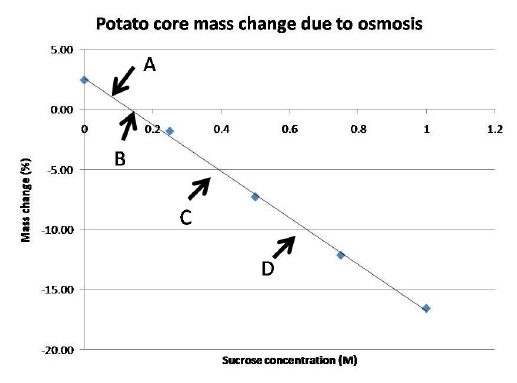
The following graph is based on potato cores soaked in various concentrations of
sucrose for twenty for house. 
|
|
|
17.
|
Using the graph above, the average mass of the potato core soaked in pure water
tends to
a. | gain mass | d. | lose water | b. | lose mass | e. | both a and c | c. | gain
water | f. | both b and
d |
|
|
|
18.
|
The potato cores in .25 M concentration of sucrose are said to be in
----.
a. | isotonic solution since they lose mass | b. | hypotonic solution since they gain
mass | c. | hypertonic solution since they lose mass | d. | saline solution
since the cores gain mass | e. | both b and d are
correct |
|
|
|
19.
|
Which of the following statement(s) is/are correct?
a. | The area marked A is referring to a hypertonic solution | b. | The area marked B is
referring to an isotonic solution | c. | The area marked C is referred to as a weak
solution | d. | The area marked D is referred to as a hypertonic solution | e. | Both B and D are
corrent |
|
Short Answer
|
|
|
Using the image below answer the following question:
|
|
|
20.
|
The image above shows a partial cell taken with TEM. By observing the diagram
carefully offer your conclusion as to what type of cell is observed (prokaryotic or eukaryotic-
animal or plant). Cite two specific examples of structure from the diagram to help support your
answer. Please feel free to label the diagram to help frame your answer.
|