Pictured:  B. Carlson, S. Contess, A. Feng, V. Oliveira, V. Wei, A. Young, K. Kolb, S. Hanchuk with Ann Stock, Ph.D. UMDNJ, Piscataway, HHMI

To see previous Pingry SMART Team projects please click here


Pingry S. M. A. R. T Team 2008-2009

Students Modeling a Research Topic

coordinated by the Center for BioMolecular Modeling, Milwaukee School of Engineering, Milwaukee, WI

Tim Herman, Ph.D, Director

Teacher Advisors- Deirdre O'Mara, Tommie Hata

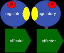
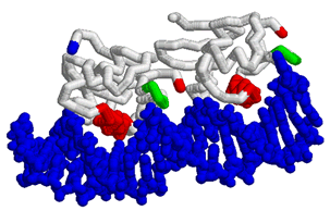
This year we are working with Anne Stock (HHMI, Center for Advanced Biotechnology, UMDNJ, Rutgers University, Piscataway, NJ) to create three dimensional models using RP-Rasmol of several proteins involved in the signal transduction pathway of bacteria. The proteins we intend to model are the second component of a two component signal transduction mechanism common in prokaryotes. The first half of the signal transduction pathway results in the phosphorylation of  a protein kinase which activates a response regulator. We intend to model this response regulator protein from various species in different states of activation. The response regulator protein consists of two domains- the regulator region and an effector region. The regulator component, when phosphorylated, will dimerize in a symetrical formation. The regulator domain, connected via flexible linker to the effector domain will bind to DNA in a winged helix motif and in a directional orientation.

The SMART Team experience begins with the selection of a mentor for our students and reading the primary literature related to the proteins of interest. With guidance from our mentor, reading the literature students create virtual three dimensional models of the proteins using RasMol. These files then are engineered for three dimensional printing on a Z-Corp machine at MSOE.

Th culmination of the project is a poster presenation at the international meeting of American Society for Biochemistry  and Molecular Biology (ASBMB) held this year in New Orleans, LA
 

Resources & Announcements:

ASBMB Meeting: Departure Sat, April 18 - return Tuesday April 21, 2009

Itinerary doc.    Letter to Parents doc.     ASBMB website

Resources:

Rasmol (better version) download to your computer

Rasmol Resource Guide- CBM, Shannon Colton, Ph.D.  Part I    Part II    Part III

SMART Teams at CBM resource page (has Rasmac link)

Dr. Stock ppt   Dr. Stock  Lab website

Protopedia Instructions:

We need to establish your proteins on Proteopedia. Please read the following instructions on how Dr. Herman would like us to follow through. You will need to request access first. Please go to the link and request accees immediately. Susan already has completed this process and can help others.

www.proteopedia.org

Tim Herman Letter for SMART Team Members regarding Protopedia

Second Letter from Tim Herman regarding Protopedia

 
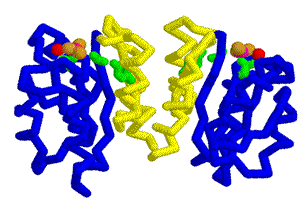
PhoB Inactive


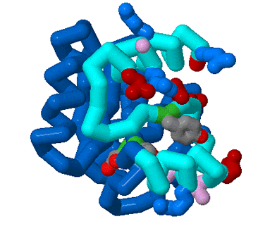
       PhoB is a protein involved in a two-component signal transduction system in prokaryotes. It is a transcription factor that activates more than 30 genes of the pho regulon. Activated under phosphate starvation conditions, PhoB is made up of two domains: the regulatory domain shown above(dimerization domain) and the effector domain (DNA binding domain).Once phosphorylated, PhoB is able to dimerize and in turn, allow binding of the effector domain to the DNA directly to pho box, where it promotes the binding of RNA polymerase.

      Only until phosphorylation will the PhoB’s inactive regulatory domain, highlighted in blue, become activated and dimerize. This dimerization occurs between two regulatory domains, interacting between the main α4β5α5 interfaces, highlighted in cyan. In PhoB’s inactive regulatory domain, there are three highly conserved and key residues, highlighted in green backbone, which are: Tyr102, Thr83, and Asp53.  Tyr102 and Thr83 are involved in the activation of inactive regulatory domain while Asp53 is a key residue for phosphorylation. Note that the same three residues also change conformations once activated, which are also highlighted in green in the active regulatory domain model. Along the dimer interface we also have numerous hydrophobic (in purple), positively charged (in blue), and negatively charged side chains (in dark red). This model also shows the carboxyl end of the regulatory protein, in red, where the linker connects it to the effector domain. Through this model we hope to assist in visualizing the before and after effects of the inactive to the active regulatory domain after phosphorylation and to understand primary step that PhoB must take in its two-component signal transduction system

Needs paper citation from Vicky


PhoB Active  Susan Contess


Winged Helix- Katherine and Ashley

Text needed here.....from K & and A

Explain the image.. what are you trying to show...whare are the colors representing.

which pdb file are you using


MtrA- Andrew and Stephanie
                                               

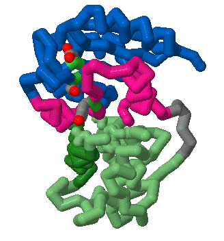
          MtrA has been isolated from species Mycobacterium tuberculosis and is a member of a typical two component signal transduction pathway consisting of a histidine kinase and response regulator. This two domain response regulator consists of an N-terminal regulatory domain and c-terminus effector domain separated by a linker region. MtrA protein is a member of the OmpR/PhoB family, which is a family of transcription factors with a winged Helix-Turn-Helix DNA binding domains (effector domain). The regulator domain is  phosphorylated by Histidine Protein Kinases and phosphorylation depends on environmental signals. Phosphorylation occurs at aspartate (#) residue near the protein’s active site found in the regulatory domain. The inactive state of the protein is associated with a conformation in which the hydroxyl group of the Thr 83 side chain points away from the active site, and the Tyr 102 residue extends outward from the surface of the regulatory domain. Conversely, in the active state, the hydroxyl group of the Thr 83 residue points toward the active site, in a position where it can interact with the phosphorylated Asp (#) residue, and the Tyr 102 residue is buried in the conformational change. These two states exist in equilibrium with phosphorylation of the regulatory domain shifting the equilibrium toward the active state. The intramolecular interface between the Regulatory domain and the effector domain in MtrA inhibits the DNA-binding activity by blocking access to recognition helix, blocking dimer formation, reducing rates of phosphorylation by trapping regulatory domain in an inactive state.

needs pdb and revision and citation


Last Updated: March 12, 2009